
X Ray Imaging
- Details
- Last Updated: Wednesday, 12 February 2014 09:58
|
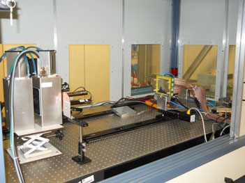
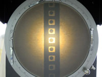
A triple GEM detector for X rays imaging has been made using the standard 10x10cm2 GEM foil produced by CERN. The detector is visible in the picture below mounted on a scan system in front of a Molybdenum X ray source. |
||
 |
 |
 |
|
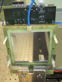
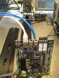
On the back side of the detector is mounted the new motherboard based on FPGA.
|
||
 |
The anode of the triple GEM detector is equipped with a row of 128 pads;
|
|
 |
The magnified image of some pads is visible on this picture alongside. |
|
|
An X ray source has been set at 30 KV with a current of 300 microampere.
|
|
|
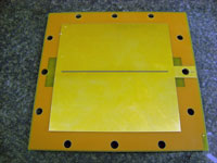
A grid realized with kapton foil and copper cladding on both side and 600 micron holes drilled on it with 2 mm pitch, Mooving the detector in front of the grid, an image is obtained as shown in this movie. Each column is acquired in 100 ms.The display dimension is 9x7 cm² |
||
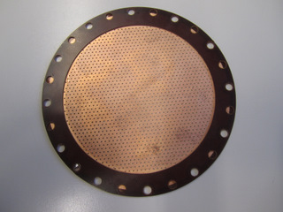 |
The grid picture |